Skip to product information

RDMS Specialty Pack: Abdomen + OB/GYN

RDMS Specialty Pack: Abdomen + OB/GYN

check_circle Digital & Printable Version

 (276+ Reviews)
Regular price €45,95
Regular price €45,95 Sale price €117,95
SAVE 61% Sold out
 

🩻 VISUAL POSITIONING GUIDES | INSTANT DIGITAL ACCESS 🎯

🔥 Instant download. Review your next projection in 60 seconds.
  • Amazon
  • American Express
  • Apple Pay
  • Bancontact
  • Diners Club
  • Discover
  • Google Pay
  • Mastercard
  • MB WAY
  • PayPal
  • Shop Pay
  • Visa
RDMS Specialty Pack: Abdomen + OB/GYN

RDMS Specialty Pack: Abdomen + OB/GYN

Regular price €45,95
Regular price €45,95 Sale price €117,95
SAVE 61% Sold out

Digital & Printable (PDF).
Note: this is a digital download

SPI done. Now see the pathology.

Specialty exams do not ask whether you read the chapter. They ask whether you can recognize the finding.

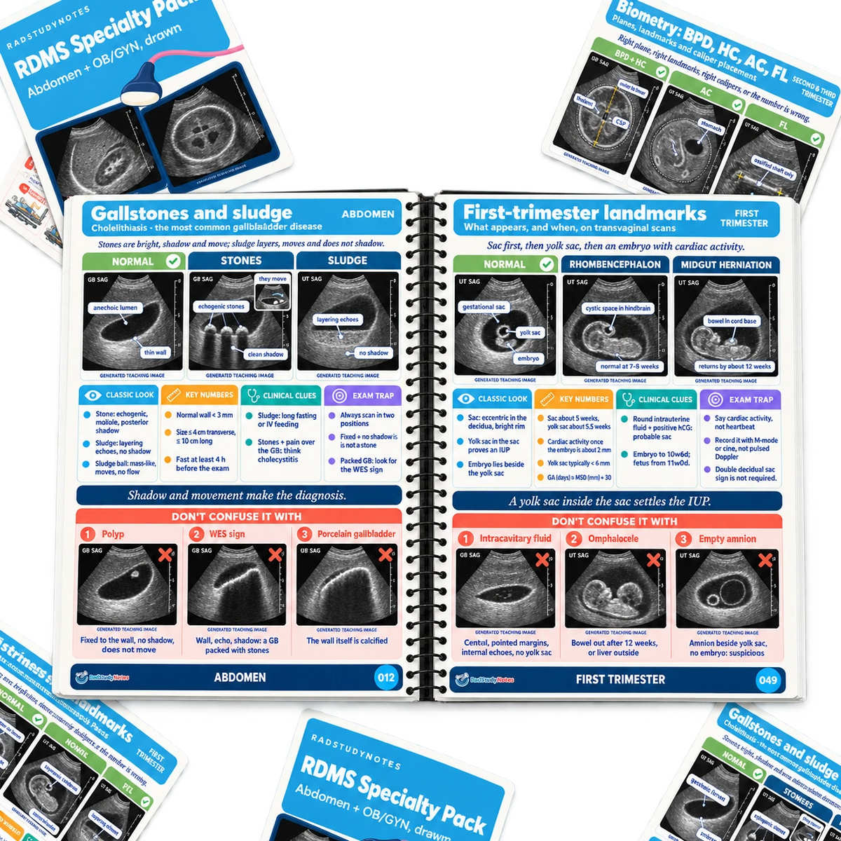
The RDMS Specialty Pack turns the Abdomen and OB/GYN content into a clear visual review: normal versus pathology side by side, the named signs drawn, the key numbers, and the look alikes that lead to the wrong answer.

Built for sonography students preparing the two specialty exams most often taken with the SPI, this 75 page guide is organized by the current ARDMS content outlines.

Open it before a specialty exam. Print it for your folder. Use it to picture the finding before the image even loads.

You are not just memorizing pathology. You are learning to tell it apart from what it is not.

  • 75 visual, print ready A4 pages

  • Abdomen: liver, gallbladder, bile ducts, pancreas, kidneys, spleen, vessels, bowel, neck and more

  • OB/GYN: normal and abnormal findings, biometry and key numbers

  • "Don't confuse it with" look alikes flagged on every topic

  • Digital PDF with instant access, in a print version and a tablet version
View full details

Our customers tell it better than we do!

Excellent 4.9 / 5
★★★★★
based on 276+ reviews
  • ★★★★★

    Clear without talking down to you

    It is simple enough to understand quickly, but it does not feel oversimplified. The image criteria and common errors are especially helpful because they show why the detail matters.

    Mia R., Second Year Radiography Student

  • ★★★★★

    The trauma pages gave me a plan

    The trauma content was one of the areas I felt least confident about. Seeing the adaptations laid out clearly helped me understand what changes and what still needs to stay in the image.

    David M., Radiography Student

  • ★★★★★

    The terminology stopped confusing me

    I kept getting caught out by different names for the same projection. Having the US and UK terminology together saved me from constantly checking multiple sources.

    Helen C., Student Radiographer

  • ★★★★★

    I wish I had this in my first semester

    I spent too long trying to turn dense notes into something I could actually remember. This is the kind of visual reference I would have wanted from the start.

    Gaby L., Radiography Student

  • ★★★★★

    Made revision feel less chaotic

    I had information everywhere, slides, screenshots, notes and textbooks. This gave me one place to review the positioning details without jumping between five different resources.

    Cindy B., Radiography Student

  • ★★★★★

    More helpful than rereading my textbook

    My textbook is useful, but it takes time to find the exact thing I need. The Atlas is what I reach for when I need to see the setup, the central ray, and the image criteria quickly.

    Tommy F., Student Radiographer

    WHAT’S INCLUDED

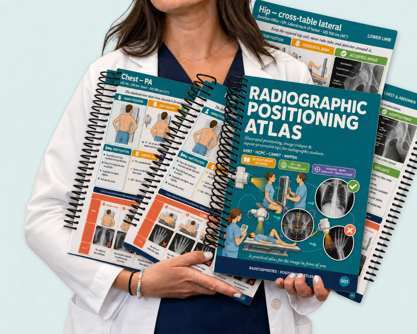
    Built for radiography students who understand the terminology in class but freeze when it is time to position a patient, place the central ray, or judge whether an image is acceptable.

    Every dense positioning concept rebuilt into visual frameworks your brain can use under pressure. No filler. Just the high yield details you need for clinicals, competencies, and exams.

    feature-item-1
    Instant Worldwide Delivery
    100% Digital delivery allows our service to be eco-friendly, affordable & versatile.
    feature-item-2
    5-Star Customer Service
    Contact us anytime
    feature-item-3
    Secure Payment
    Our checkout accepts all Visa, Mastercard, as well as PayPal, GooglePay, Apple Pay, Klarna & many more.
    feature-item-4
    Focused Visual Study Support
    Includes diagrams, memory cues, and structured summaries of key topics.

    Built for radiography students who know that memorising the theory is not enough when a real patient is in front of you.

    Excellent 4.9 / 5

    • I stopped panicking before clinicals 😅

      I knew the terminology, but I still felt anxious before clinical days because I never knew what detail I was forgetting. The visual layout helped me review the whole projection without getting lost in my notes.

      Kate S., Second Year Radiography Student

    • The central ray finally made sense

      I had memorised so many positions but could not always connect the central ray to what the image needed to show. Having the setup and evaluation points on the same page made a huge difference

      John M., Radiography Student

    • It showed me what my notes were missing

      My lecture slides explained the theory, but they did not help me picture the projection in front of me. This gave me the visual connection I was missing.

      Reana R., Student Radiographer

    • The image checks are gold

      The rotation, centering, collimation and exposure checks gave me a much better way to approach image critique. I feel like I can now explain what I am looking for instead of just guessing.

      Lara D., First Year Radiography Student

    • Useful when I only have ten minutes

      I use it before a lecture, between clinical days, or when I need a quick refresher before revision. It is much easier to return to than trying to search through pages of handwritten notes.

      Ema S., Radiography Student

      Product Specification and FAQ

      Product Specification

      Format: High resolution digital PDF, 76 A4 pages

      Content: Visual radiographic positioning guidance covering fundamentals, chest and abdomen, upper limb, lower limb, spine, skull and facial bones, trauma, and quick reference tools

      Study Tools: Patient positioning diagrams, central ray guidance, collimation cues, image evaluation criteria, common errors, angle tables, and a repeat or accept decision tree

      Organization: Color coded visual frameworks built to make dense positioning concepts easier to understand and recall

      Use Case: Supplemental personal study resource for lectures, clinicals, competencies, and exam revision

      Access: Instant digital access on iPad, tablet, desktop, or smartphone. Print at home for offline review

      Shipping Information

      Shipping Information

      • Delivery Method: Digital download via email
      • Processing Time: Receive your product immediately after purchase
      • Access: Instant access to the PDF file upon delivery
      • Support: Contact us if you encounter any issues with the download

      Can I print the study guide?

      Yes, the PDF is printable. You can print it out for physical reference or use it digitally on your devices.

      Are there any updates or additional materials included?

      The PDF you receive is the complete study guide. However, if there are any updates or additional materials, you will be notified via email.

      Who is it for?

      It's designed for visual learners and busy professionals who want a faster, clearer way to review the densest scientific foundations chapters. It's most useful as a complement to your main study materials — the visual review layer most learners are missing when they go back to the textbook.

      Still have questions?

      You can reach us anytime at info.radstudynotes@gmail.com for support.