

STOP SECOND GUESSING. START SEEING THE WHOLE PROJECTION.
Patient position. Central ray. Collimation. Image criteria. See every critical decision connected on one visual page.
Built for radiography students preparing for ARRT, CAMRT, university exams, clinical placements and competency checks worldwide.


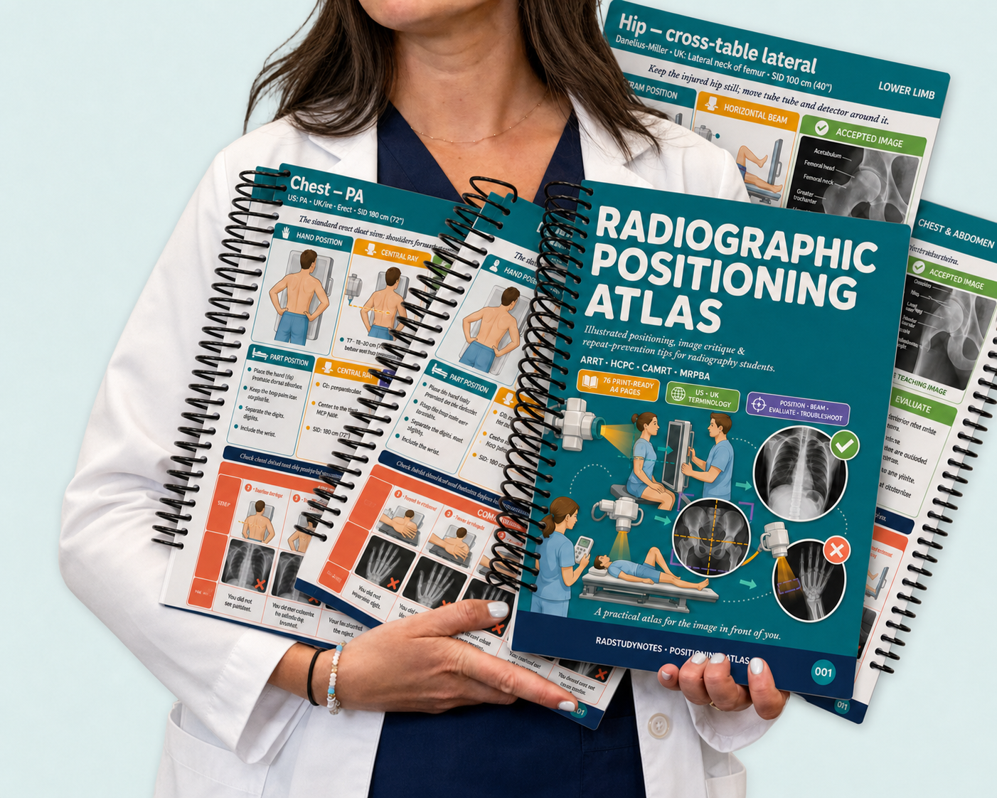
Radiographic Positioning Atlas
Radiographic Positioning Atlas
Digital & Printable Version
2026 Edition. You can know the terminology and still freeze when an exam question or clinical scenario asks what comes next.Dense notes force you to assemble the entire projection in your head when the pressure is already on.
This Atlas puts patient setup, central ray, collimation, accepted image criteria and common errors together, so you can understand the complete projection faster and recall it when it counts.
One international visual system for ARRT review, CAMRT preparation, university exams and clinical competencies.
🩻 See the complete projection at a glance
🎯 Review positioning, procedures and image evaluation for exams and competencies
🌍 US and UK terminology for radiography students worldwide
📚 76 visual A4 pages designed for faster, more focused recall
📱 Instant access. Study on your phone, tablet, laptop or printed pages
🩻 ARRT REVIEW | CAMRT PREP | CLINICALS | COMPETENCIES 🎯
Couldn't load pickup availability

Radiographic Positioning Atlas


RADIOGRAPHY STUDENTS SAY IT BEST
-

★★★★★
The fastest way I found to review positioning
I still use my textbook, but I was wasting so much time searching through chapters for one projection. This gives me the setup, central ray and image criteria in seconds.
-

★★★★★
Finally, terminology that works internationally 🌍
Our course uses UK terminology, while many online resources use US terms. Having both in one place has stopped a lot of unnecessary confusion.
-

★★★★★
The trauma section gave me an actual plan
Trauma positioning was one of the areas I kept avoiding. The adaptations are laid out clearly without making the pages feel overwhelming.
-

★★★★★
Made my CAMRT review feel much more organized
I had notes scattered across lectures, screenshots and textbooks. The Atlas helped me bring the positioning content together and see what I still needed to review.


WHAT’S INCLUDED
76 visual A4 pages. 8 focused sections. Every major body region, plus trauma adaptations and quick reference tools.
See the setup. Know where the central ray goes. Understand what the final image should show. Catch the mistakes that lead to repeats.
No scattered screenshots. No pages of filler. Just the positioning details you need for ARRT review, CAMRT preparation, university exams, clinical placements and competencies.

📐 Positioning Fundamentals
The language of positioning
· Body positions and planes
· Central ray, SID and divergence
· The four image checks
· Image evaluation criteria
· Radiation protection in practice
🫁 Chest & Abdomen
Chest PA, lateral, portable, decubitus and apical views
· Ribs above and below the diaphragm
· Abdomen supine and erect projections
🖐️ Upper Limb
Hand, fingers, wrist and scaphoid
· Forearm, elbow and humerus
· Shoulder, scapula, clavicle and AC joints
· The views students second guess most
🦵 Lower Limb & Pelvis
Foot, ankle and mortise
· Tibia and fibula
· Knee, patella and femur
· Hip, pelvis and cross table lateral positioning
🦴 Spine
Cervical, thoracic and lumbar spine
· Odontoid and swimmer’s lateral
· Obliques, cross table views, sacrum and coccyx
🧠 Skull & Facial Bones
Skull projections
· Facial bones and orbits
· Nasal bones · Mandible and TMJ
· Paranasal sinuses
🚑 Trauma & Clinical Adaptations
The principles of trauma positioning
· Upper limb, lower limb and spine trauma views
· Paediatric and bariatric adaptations
⚡ Quick Reference Tools
Repeat or accept decision tree
· Complete angle table
· SID and FRD table
· US and UK terminology guide
· Fast index


THE EXAM DOES NOT ASK WHETHER YOU READ THE CHAPTER.
It asks whether you can apply it.
Across ARRT review, CAMRT preparation, university exams and clinical competencies, you need to connect patient position, central ray, collimation, anatomy demonstrated and image evaluation.
Your textbooks often teach those pieces across different pages and chapters.
The Atlas brings them together in one visual workflow, so you can review faster, build stronger recall and stop guessing when the answer, the competency or the patient is in front of you.
See the setup. Understand the image. Recall it when it counts.

YOU DO NOT NEED ANOTHER PILE OF NOTES.
You need patient position, central ray, collimation, image criteria and common errors to click together.
Open one page and see the entire projection.
Less searching. Less second guessing. More clarity when it matters.
Built for radiography students worldwide who refuse to let dense notes decide how confident they feel in exams or clinicals.
Excellent 4.9 / 5

Radiographic Positioning Atlas
Radiographic Positioning Atlas
Product Specification and FAQ
Product Specification
Product Specification
Format: High resolution digital PDF, 76 A4 pages
Content: Visual radiographic positioning guidance covering fundamentals, chest and abdomen, upper limb, lower limb, spine, skull and facial bones, trauma, and quick reference tools
Study Tools: Patient positioning diagrams, central ray guidance, collimation cues, image evaluation criteria, common errors, angle tables, and a repeat or accept decision tree
Organization: Color coded visual frameworks built to make dense positioning concepts easier to understand and recall
Use Case: Supplemental personal study resource for lectures, clinicals, competencies, and exam revision
Access: Instant digital access on iPad, tablet, desktop, or smartphone. Print at home for offline review
Deivery & Access
Deivery & Access
Delivery Method:Instant digital download sent by email
Delivery Time: Access the complete Atlas immediately after purchase
Devices: Open the PDF on your phone, tablet, laptop or desktop
Support: Contact us if you experience any problem accessing your files
Can I print the study guide?
Can I print the study guide?
Yes, the PDF is printable. You can print it out for physical reference or use it digitally on your devices.
Can this help me prepare for ARRT, CAMRT and university exams?
Can this help me prepare for ARRT, CAMRT and university exams?
Yes. The Atlas supports focused review of radiographic positioning, procedures, anatomy and image evaluation.
These areas are relevant to ARRT Radiography review, CAMRT certification preparation, university assessments and clinical competencies.
It is not a complete official exam course. It is the visual positioning resource you use alongside your official exam blueprint, program materials and textbooks.
Who is it for?
Who is it for?
The Atlas is built for radiography and radiologic technology students who want positioning to feel clearer, faster to review and easier to recall.
It is especially useful for visual learners, ARRT candidates, CAMRT candidates, UK student radiographers, internationally educated technologists and anyone preparing for university exams, clinical placement or competency assessments.
Still have questions?
Still have questions?
You can reach us anytime at info.radstudynotes@gmail.com for support.